Vasectomy Reversal: In Pictures
What does a vasectomy reversal look like?
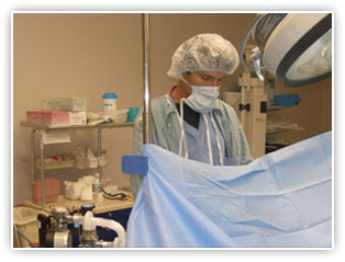
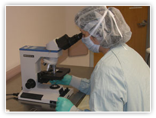
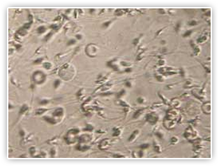
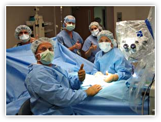
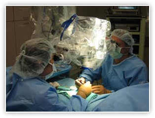
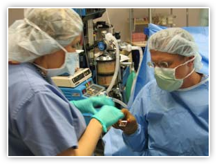
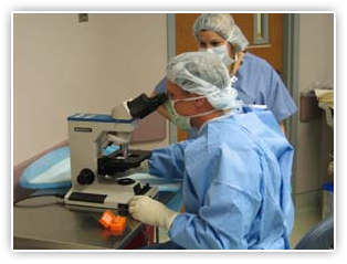
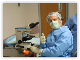
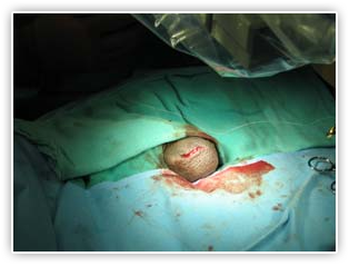
Have you ever wondered what a vasectomy reversal procedure looks like under the microscope? Follow Dr. Bastuba into the operating room and look over his shoulder to view a microsurgical vasectomy reversal pictures.
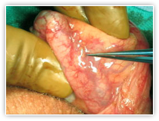
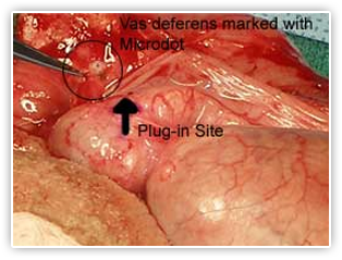
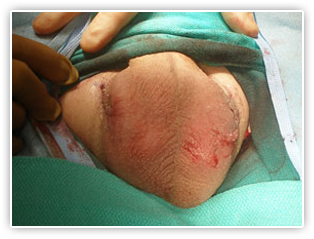
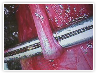
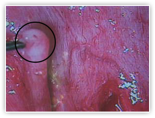
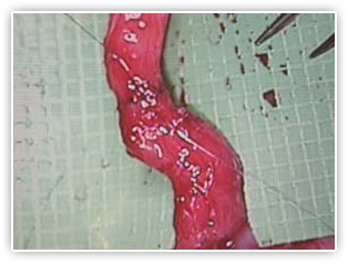
Vasovasostomy
The vasovasostomy is the most commonly performed vasectomy reversal surgery. During a vasovasostomy, the separated ends of the vas deferens are reconnected. See Dr. Bastuba perform a vasovasostomy with these step-by-step reverse vasectomy pictures.
![]()
![]()
Many of our patients are very interested in seeing an actual vasectomy reversal procedure. Others are uncomfortable seeing detailed photographs and prefer not to know all of the particulars. If you are ready to get started, please use the buttons below to navigate our slideshow.

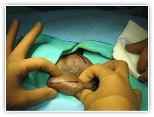
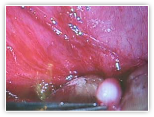
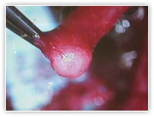
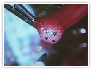

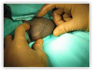
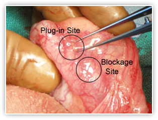
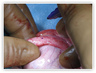
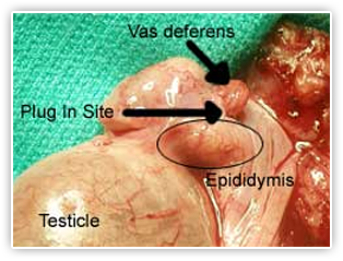
Vasoepididymostomy (VE)
During a vasectomy reversal it may turn out that the more complicated reversal procedure, a vasoepididymostomy (VE, also known as epididymovasostomy), will be necessary. See Dr. Bastuba perform a vasoepididymostomy step-by-step.
![]()
![]()
Many of our patients are very interested in seeing an actual vasectomy reversal procedure. Others are uncomfortable seeing detailed photographs and prefer not to know all of the particulars. If you are ready to get started, please use the buttons below to navigate our slideshow.